Functional Assessment of Cerebral Capillaries using Single Capillary Reporters in Ultrasound Localization Microscopy

0

Sign in to get full access
Overview
- This paper presents a new technique called Ultrasound Localization Microscopy (ULM) that can assess the function of individual cerebral capillaries, which are the smallest blood vessels in the brain.
- The researchers developed a method to label specific capillaries with reporter molecules that emit ultrasound signals, allowing them to track the behavior of individual capillaries.
- This approach provides a way to study the dynamics of blood flow and oxygen delivery at the capillary level, which is crucial for understanding brain function and dysfunction.
Plain English Explanation
The brain relies on a complex network of tiny blood vessels called capillaries to deliver oxygen and nutrients. Towards Transcranial 3D Ultrasound Localization Microscopy in Non-human Primates shows that the function of these individual capillaries can be measured using a new imaging technique called Ultrasound Localization Microscopy (ULM).
The researchers developed a way to label specific capillaries with special reporter molecules that emit ultrasound signals. This allows them to track the behavior of individual capillaries, such as how blood flows through them and how much oxygen is delivered. By studying the dynamics of capillary function, scientists can gain important insights into brain health and disease.
For example, Framework to Generate Perfusion Map from CT shows how capillary-level imaging could help identify problems with blood flow that contribute to conditions like stroke or Alzheimer's disease. The ability to assess capillary function at this microscopic level represents an important advance in our understanding of the brain's vascular system.
Technical Explanation
The key innovation in this paper is the use of "single capillary reporters" - molecules that can be selectively attached to individual capillaries and emit ultrasound signals. By detecting these signals, the researchers were able to create high-resolution maps of blood flow and oxygen delivery at the capillary level using Ultrasound Localization Microscopy (ULM).
The experimental setup involved injecting the reporter molecules into the bloodstream of mice and then using a specialized ultrasound imaging system to detect the signals from individual capillaries. Neurovascular Segmentation from SOCT with Deep Learning and Synthetic Training shows how deep learning can be used to enhance the analysis of these types of microscopic vascular images.
The researchers were able to track the dynamics of blood flow and oxygen levels within specific capillaries over time. This allowed them to assess the functional state of the capillary network and identify any regions with impaired blood supply or oxygenation. Enhancing Super-Resolution Ultrasound Localisation through Multi-Frame Processing describes how advanced signal processing techniques can further improve the resolution and accuracy of ULM imaging.
Critical Analysis
The use of single capillary reporters and ULM imaging represents a significant advance in our ability to study the microscopic functioning of the brain's vascular system. However, the technique does have some limitations:
- The reporter molecules must be injected into the bloodstream, which may not be feasible for all applications, especially in humans.
- The current imaging setup requires the skull to be surgically exposed, limiting its use to preclinical animal studies. Inverse Problem Approach to Aberration Correction In Vivo discusses some of the challenges in applying these techniques transcranially.
- The technique provides a snapshot of capillary function at a given moment, rather than continuous long-term monitoring, which would be valuable for understanding dynamic changes in brain physiology.
Future research should focus on addressing these limitations, such as developing less invasive reporter molecules or improving the penetration of ultrasound signals through the skull. Broader adoption of this technology could lead to important breakthroughs in our understanding and treatment of neurological disorders.
Conclusion
This paper presents a novel approach to imaging the functional state of individual cerebral capillaries using Ultrasound Localization Microscopy and custom-designed reporter molecules. By tracking the dynamics of blood flow and oxygen delivery at the capillary level, researchers can gain unprecedented insights into the vascular underpinnings of brain function and dysfunction.
While the current implementation has some practical limitations, the potential of this technology to transform our understanding of the brain's vascular system is substantial. Further development of these techniques could lead to new diagnostic and therapeutic approaches for a wide range of neurological conditions.
This summary was produced with help from an AI and may contain inaccuracies - check out the links to read the original source documents!
Related Papers
0
Functional Assessment of Cerebral Capillaries using Single Capillary Reporters in Ultrasound Localization Microscopy
Stephen A Lee, Alexis Leconte, Alice Wu, Joshua Kinugasa, Jonathan Poree, Andreas Linninger, Jean Provost
The brain's microvascular cerebral capillary network plays a vital role in maintaining neuronal health, yet capillary dynamics are still not well understood due to limitations in existing imaging techniques. Here, we present Single Capillary Reporters (SCaRe) for transcranial Ultrasound Localization Microscopy (ULM), a novel approach enabling non-invasive, whole-brain mapping of single capillaries and estimates of their transit-time as a neurovascular biomarker. We accomplish this first through computational Monte Carlo and ultrasound simulations of microbubbles flowing through a fully-connected capillary network. We unveil distinct capillary flow behaviors which informs methodological changes to ULM acquisitions to better capture capillaries in vivo. Subsequently, applying SCaRe-ULM in vivo, we achieve unprecedented visualization of single capillary tracks across brain regions, analysis of layer-specific capillary heterogeneous transit times (CHT), and characterization of whole microbubble trajectories from arterioles to venules. Lastly, we evaluate capillary biomarkers using injected lipopolysaccharide to induce systemic neuroinflammation and track the increase in SCaRe-ULM CHT, demonstrating the capability to detect subtle capillary functional changes. SCaRe-ULM represents a significant advance in studying microvascular dynamics, offering novel avenues for investigating capillary patterns in neurological disorders and potential diagnostic applications.
Read more7/12/2024

0
Towards Transcranial 3D Ultrasound Localization Microscopy of the Nonhuman Primate Brain
Paul Xing, Vincent Perrot, Adan Ulises Dominguez-Vargas, Stephan Quessy, Numa Dancause, Jean Provost
Hemodynamic changes occur in stroke and neurodegenerative diseases. Developing imaging techniques allowing the in vivo visualization and quantification of cerebral blood flow would help better understand the underlying mechanism of those cerebrovascular diseases. 3D ultrasound localization microscopy (ULM) is a novel technology that can map the microvasculature of the brain at large depth and has been mainly used until now in rodents. Here, we demonstrated the feasibility of 3D ULM of the nonhuman primate (NHP) brain with a single 256-channels programmable ultrasound scanner. We achieved a highly resolved vascular map of the macaque brain at large depth in presence of craniotomy and durectomy using an 8-MHz multiplexed matrix probe. We were able to distinguish vessels as small as 26.9 {mu}m. We also demonstrated that transcranial imaging of the macaque brain at similar depth was feasible using a 3-MHz probe and achieved a resolution of 60.4 {mu}m. This work paves the way to clinical application of 3D ULM.
Read more4/5/2024
👨🏫
0
Correlating Stroke Risk with Non-Invasive Tracing of Brain Blood Dynamic via a Portable Speckle Contrast Optical Spectroscopy Laser Device
Yu Xi Huang, Simon Mahler, Aidin Abedi, Julian Michael Tyszka, Yu Tung Lo, Patrick D. Lyden, Jonathan Russin, Charles Liu, Changhuei Yang
Stroke poses a significant global health threat, with millions affected annually, leading to substantial morbidity and mortality. Current stroke risk assessment for the general population relies on markers such as demographics, blood tests, and comorbidities. A minimally invasive, clinically scalable, and cost-effective way to directly measure cerebral blood flow presents an opportunity. This opportunity has potential to positively impact effective stroke risk assessment prevention and intervention. Physiological changes in the cerebral vascular system, particularly in response to carbon dioxide level changes and oxygen deprivation, such as during breath-holding, can offer insights into stroke risk assessment. However, existing methods for measuring cerebral perfusion reserve, such as blood flow and blood volume changes, are limited by either invasiveness or impracticality. Here, we propose a transcranial approach using speckle contrast optical spectroscopy (SCOS) to non-invasively monitor regional changes in brain blood flow and volume during breath-holding. Our study, conducted on 50 individuals classified into two groups (low-risk and higher-risk for stroke), shows significant differences in blood dynamic changes during breath-holding between the two groups, providing physiological insights for stroke risk assessment using a non-invasive quantification paradigm. Given its cost-effectiveness, scalability, portability, and simplicity, this laser-centric tool has significant potential in enhancing the pre-screening of stroke and mitigating strokes in the general population through early diagnosis and intervention.
Read more7/24/2024
0
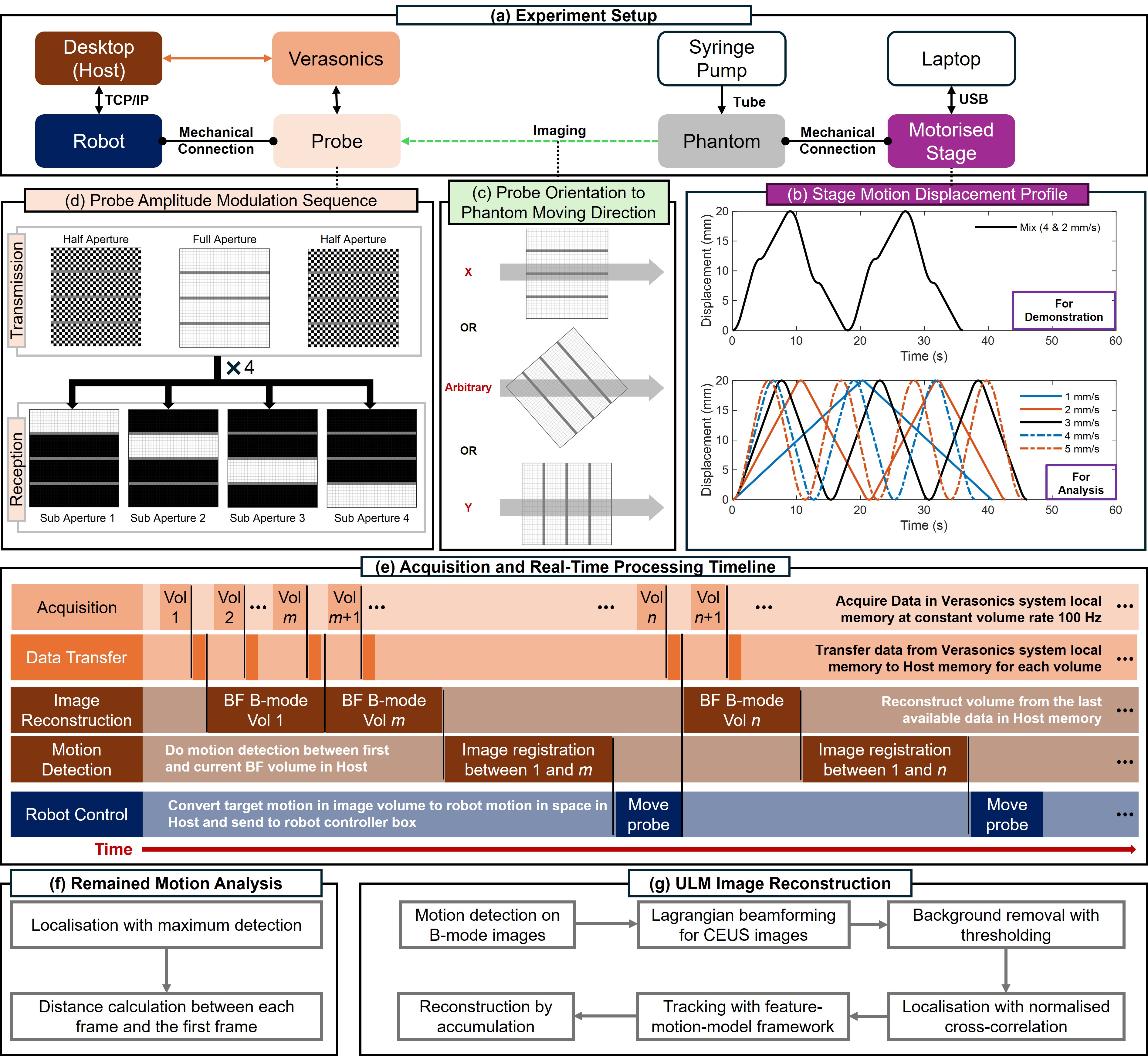
Online 4D Ultrasound-Guided Robotic Tracking Enables 3D Ultrasound Localisation Microscopy with Large Tissue Displacements
Jipeng Yan, Shusei Kawara, Qingyuan Tan, Jingwen Zhu, Bingxue Wang, Matthieu Toulemonde, Honghai Liu, Ying Tan, Meng-Xing Tang
Super-Resolution Ultrasound (SRUS) imaging through localising and tracking microbubbles, also known as Ultrasound Localisation Microscopy (ULM), has demonstrated significant potential for reconstructing microvasculature and flows with sub-diffraction resolution in clinical diagnostics. However, imaging organs with large tissue movements, such as those caused by respiration, presents substantial challenges. Existing methods often require breath holding to maintain accumulation accuracy, which limits data acquisition time and ULM image saturation. To improve image quality in the presence of large tissue movements, this study introduces an approach integrating high-frame-rate ultrasound with online precise robotic probe control. Tested on a microvasculature phantom with translation motions up to 20 mm, twice the aperture size of the matrix array used, our method achieved real-time tracking of the moving phantom and imaging volume rate at 85 Hz, keeping majority of the target volume in the imaging field of view. ULM images of the moving cross channels in the phantom were successfully reconstructed in post-processing, demonstrating the feasibility of super-resolution imaging under large tissue motions. This represents a significant step towards ULM imaging of organs with large motion.
Read more9/18/2024