NeuroMorphix: A Novel Brain MRI Asymmetry-specific Feature Construction Approach For Seizure Recurrence Prediction

0

Sign in to get full access
Overview
- Novel brain MRI asymmetry-specific feature construction approach called "NeuroMorphix" for predicting seizure recurrence
- Combines brain MRI data with machine learning to identify unique brain patterns linked to seizure risk
- Aims to improve seizure prediction and personalized treatment for epilepsy patients
Plain English Explanation
The paper presents a new approach called "NeuroMorphix" that uses brain MRI scans to predict the risk of seizure recurrence in epilepsy patients. Epilepsy is a neurological condition characterized by recurrent seizures, and being able to accurately predict seizure risk is important for providing the right treatment.
NeuroMorphix works by looking for unique patterns or "features" in the brain MRI scans that are associated with a higher likelihood of future seizures. These features are based on subtle differences, or "asymmetries," between the left and right sides of the brain. The researchers developed a specialized algorithm to identify these brain asymmetries and use them to train a machine learning model to predict seizure risk.
By using this brain-specific approach, the researchers hope to improve upon existing seizure prediction methods that may rely more on general clinical factors. The goal is to provide more personalized and effective care for epilepsy patients, potentially helping them better manage their condition and reduce the impact of seizures on their daily lives.
Technical Explanation
The NeuroMorphix approach involves several key steps:
-
Preprocessing brain MRI data: The researchers first preprocessed the brain MRI scans to segment the brain into distinct regions and align the left and right hemispheres.
-
Extracting brain asymmetry features: They then developed a novel algorithm to quantify the degree of asymmetry between corresponding regions in the left and right brain hemispheres. This resulted in a set of brain-specific features that capture the unique asymmetry patterns for each patient.
-
Training a machine learning model: The asymmetry features were used to train a machine learning model, specifically a random forest classifier, to predict the risk of seizure recurrence for each patient.
-
Evaluating model performance: The researchers tested the NeuroMorphix model on a dataset of epilepsy patients and compared its performance to other seizure prediction approaches. They found that the brain asymmetry-specific features improved the model's ability to accurately predict seizure risk.
Critical Analysis
The NeuroMorphix approach offers a novel and potentially valuable way to leverage brain MRI data for seizure prediction in epilepsy patients. By focusing on brain asymmetries, the researchers have identified a unique set of features that may provide insights into the underlying neurological mechanisms of seizures.
However, the study has some limitations. The sample size was relatively small, and the model was tested on a single dataset. Further research is needed to validate the approach on larger, more diverse patient populations and to compare it to other state-of-the-art seizure prediction methods.
Additionally, the paper does not delve into the interpretability of the NeuroMorphix model. Understanding how the identified brain asymmetry features relate to seizure risk could be crucial for translating the research into clinical practice and helping physicians make more informed treatment decisions.
Conclusion
The NeuroMorphix approach represents a promising step towards improving seizure prediction and personalized treatment for epilepsy patients. By leveraging brain-specific features based on MRI asymmetries, the researchers have developed a novel machine learning-based tool that could enhance our understanding of the neurological underpinnings of seizures and lead to more effective management strategies for this debilitating condition.
This summary was produced with help from an AI and may contain inaccuracies - check out the links to read the original source documents!
Related Papers
0
NeuroMorphix: A Novel Brain MRI Asymmetry-specific Feature Construction Approach For Seizure Recurrence Prediction
Soumen Ghosh, Viktor Vegh, Shahrzad Moinian, Hamed Moradi, Alice-Ann Sullivan, John Phamnguyen, David Reutens
Seizure recurrence is an important concern after an initial unprovoked seizure; without drug treatment, it occurs within 2 years in 40-50% of cases. The decision to treat currently relies on predictors of seizure recurrence risk that are inaccurate, resulting in unnecessary, possibly harmful, treatment in some patients and potentially preventable seizures in others. Because of the link between brain lesions and seizure recurrence, we developed a recurrence prediction tool using machine learning and clinical 3T brain MRI. We developed NeuroMorphix, a feature construction approach based on MRI brain anatomy. Each of seven NeuroMorphix features measures the absolute or relative difference between corresponding regions in each cerebral hemisphere. FreeSurfer was used to segment brain regions and to generate values for morphometric parameters (8 for each cortical region and 5 for each subcortical region). The parameters were then mapped to whole brain NeuroMorphix features, yielding a total of 91 features per subject. Features were generated for a first seizure patient cohort (n = 169) categorised into seizure recurrence and non-recurrence subgroups. State-of-the-art classification algorithms were trained and tested using NeuroMorphix features to predict seizure recurrence. Classification models using the top 5 features, ranked by sequential forward selection, demonstrated excellent performance in predicting seizure recurrence, with area under the ROC curve of 88-93%, accuracy of 83-89%, and F1 score of 83-90%. Highly ranked features aligned with structural alterations known to be associated with epilepsy. This study highlights the potential for targeted, data-driven approaches to aid clinical decision-making in brain disorders.
Read more4/17/2024
🔗
0
Towards robust radiomics and radiogenomics predictive models for brain tumor characterization
Maria Nadeem, Asma Shaheen, Muhammad F. A. Chaudhary, Hassan Mohy-ud-Din
In the context of brain tumor characterization, we focused on two key questions: (a) stability of radiomics features to variability in multiregional segmentation masks obtained with fully-automatic deep segmentation methods and (b) subsequent impact on predictive performance on downstream tasks: IDH prediction and Overall Survival (OS) classification. We further constrained our study to limited computational resources setting which are found in underprivileged, remote, and (or) resource-starved clinical sites in developing countries. We employed seven SOTA CNNs which can be trained with limited computational resources and have demonstrated superior segmentation performance on BraTS challenge. Subsequent selection of discriminatory features was done with RFE-SVM and MRMR. Our study revealed that highly stable radiomics features were: (1) predominantly texture features (79.1%), (2) mainly extracted from WT region (96.1%), and (3) largely representing T1Gd (35.9%) and T1 (28%) sequences. Shape features and radiomics features extracted from the ENC subregion had the lowest average stability. Stability filtering minimized non-physiological variability in predictive models as indicated by an order-of-magnitude decrease in the relative standard deviation of AUCs. The non-physiological variability is attributed to variability in multiregional segmentation maps obtained with fully-automatic CNNs. Stability filtering significantly improved predictive performance on the two downstream tasks substantiating the inevitability of learning novel radiomics and radiogenomics models with stable discriminatory features. The study (implicitly) demonstrates the importance of suboptimal deep segmentation networks which can be exploited as auxiliary networks for subsequent identification of radiomics features stable to variability in automatically generated multiregional segmentation maps.
Read more6/12/2024
🏷️
0
Brain Tumor Classification From MRI Images Using Machine Learning
Vidhyapriya Ranganathan, Celshiya Udaiyar, Jaisree Jayanth, Meghaa P V, Srija B, Uthra S
Brain tumor is a life-threatening problem and hampers the normal functioning of the human body. The average five-year relative survival rate for malignant brain tumors is 35.6 percent. For proper diagnosis and efficient treatment planning, it is necessary to detect the brain tumor in early stages. Due to advancement in medical imaging technology, the brain images are taken in different modalities. The ability to extract relevant characteristics from magnetic resonance imaging (MRI) scans is a crucial step for brain tumor classifiers. Several studies have proposed various strategies to extract relevant features from different modalities of MRI to predict the growth of abnormal tumors. Most techniques used conventional methods of image processing for feature extraction and machine learning for classification. More recently, the use of deep learning algorithms in medical imaging has resulted in significant improvements in the classification and diagnosis of brain tumors. Since tumors are located at different regions of the brain, localizing the tumor and classifying it to a particular category is a challenging task. The objective of this project is to develop a predictive system for brain tumor detection using machine learning(ensembling).
Read more7/16/2024
0
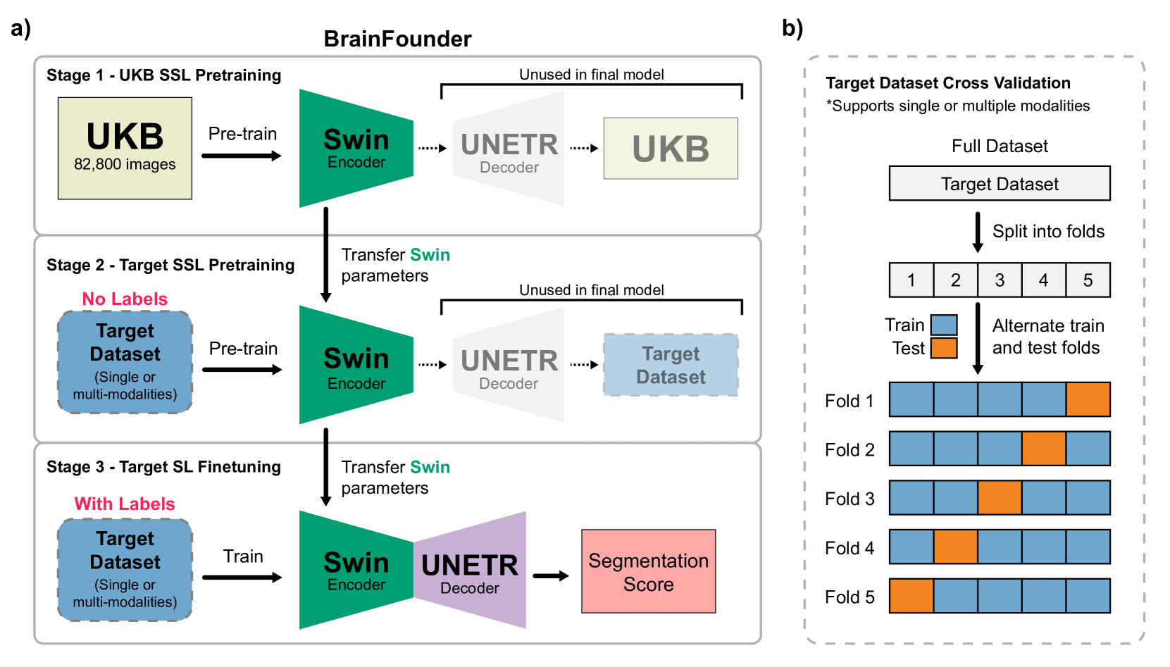
BrainFounder: Towards Brain Foundation Models for Neuroimage Analysis
Joseph Cox, Peng Liu, Skylar E. Stolte, Yunchao Yang, Kang Liu, Kyle B. See, Huiwen Ju, Ruogu Fang
The burgeoning field of brain health research increasingly leverages artificial intelligence (AI) to interpret and analyze neurological data. This study introduces a novel approach towards the creation of medical foundation models by integrating a large-scale multi-modal magnetic resonance imaging (MRI) dataset derived from 41,400 participants in its own. Our method involves a novel two-stage pretraining approach using vision transformers. The first stage is dedicated to encoding anatomical structures in generally healthy brains, identifying key features such as shapes and sizes of different brain regions. The second stage concentrates on spatial information, encompassing aspects like location and the relative positioning of brain structures. We rigorously evaluate our model, BrainFounder, using the Brain Tumor Segmentation (BraTS) challenge and Anatomical Tracings of Lesions After Stroke v2.0 (ATLAS v2.0) datasets. BrainFounder demonstrates a significant performance gain, surpassing the achievements of the previous winning solutions using fully supervised learning. Our findings underscore the impact of scaling up both the complexity of the model and the volume of unlabeled training data derived from generally healthy brains, which enhances the accuracy and predictive capabilities of the model in complex neuroimaging tasks with MRI. The implications of this research provide transformative insights and practical applications in healthcare and make substantial steps towards the creation of foundation models for Medical AI. Our pretrained models and training code can be found at https://github.com/lab-smile/GatorBrain.
Read more8/14/2024